

When you visit your dentist for a routine check-up or a specific dental issue, one of the essential tools they use to assess your oral health is dental X-rays. These diagnostic images may seem like a routine part of the dental experience, but they play a crucial role in helping your dentist identify and address dental problems effectively. In this blog post, we’ll explore why dental X-rays are so important in maintaining your oral health.
Understanding Dental X-Rays
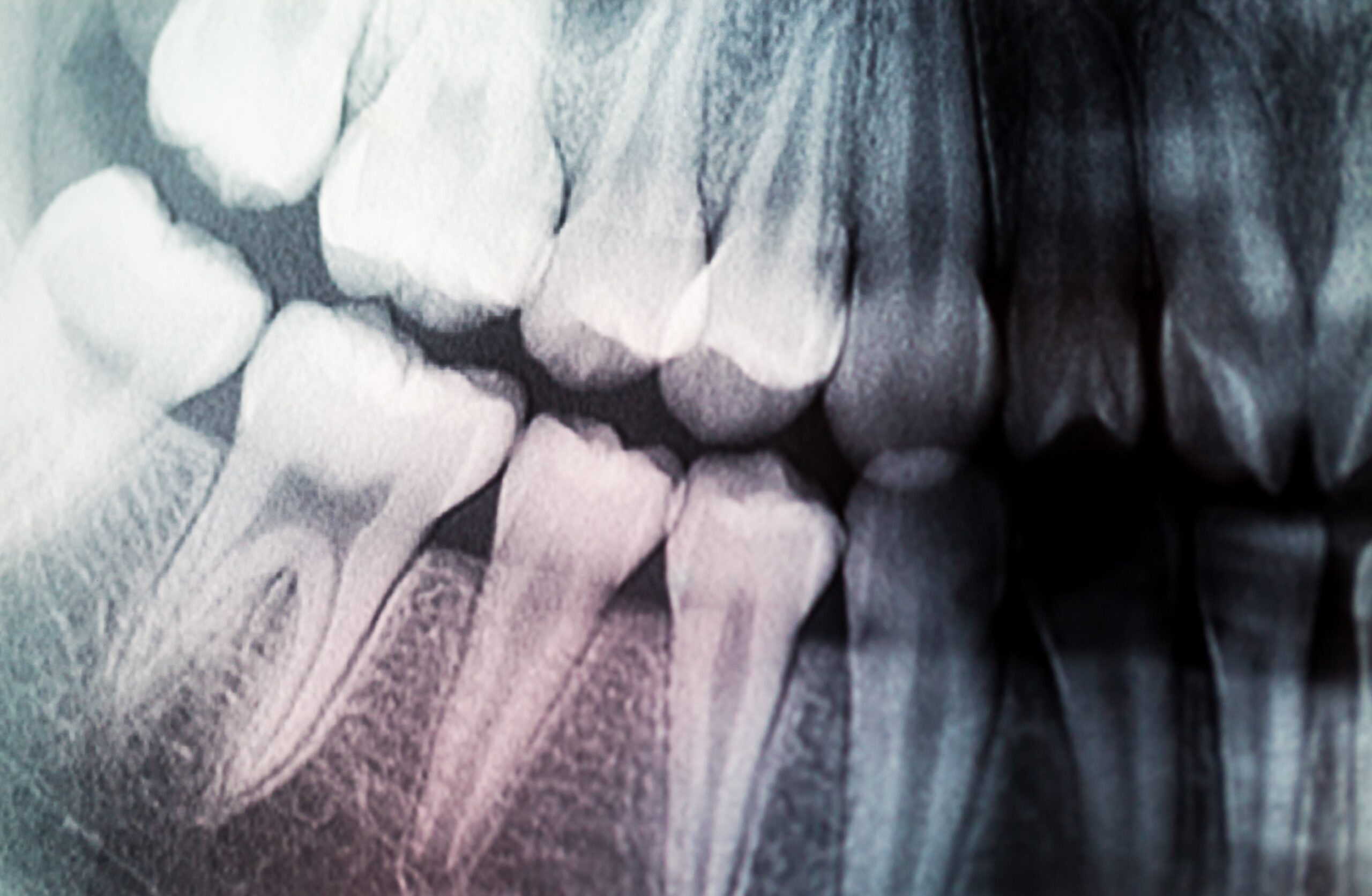
Dental X-rays, also known as radiographs, are diagnostic images of the teeth, gums, jawbone, and surrounding oral structures. These images are created using a specialized X-ray machine that emits a small amount of ionizing radiation to capture detailed pictures of your oral anatomy.
Dental X-rays are safe when administered by trained professionals, and modern equipment minimizes radiation exposure. Dentists take precautions, such as using lead aprons and thyroid collars, to ensure patient safety during the procedure. The benefits of early diagnosis and effective treatment that dental X-rays offer often outweigh the minimal risks associated with radiation exposure.
Types of dental X-rays include:
| Type of Dental X-ray | Purpose | Process | Area Covered |
| Bitewing X-rays | Detects cavities between teeth, and assess gum disease. | Patient bites down on a paper holder to position the film or sensor. | Focuses on the crowns of the back teeth. |
| Periapical X-rays | Provide a full view of one or two teeth, from crown to root. | Film or sensor is placed beneath the tooth to capture the entire tooth and surrounding bone. | Useful for detecting issues below the gum line, like impacted teeth, abscesses, cysts, and bone changes. |
| Panoramic X-rays | Offer a broad view of the entire dental arch, jaws, and sometimes sinuses and jaw joints. | Machine rotates around the patient’s head for a full arc of images. | Ideal for orthodontic planning, assessing impacted teeth, detecting jaw disorders, and comprehensive teeth and bone assessment. |
| Occlusal X-rays | View large areas of the upper or lower jaw. | Film is placed between the open jaws to capture a full view of the dental arch. | Detects extra teeth, jaw fractures, cleft palate, cysts, or growths. |
| Cephalometric X-rays | Show an entire side of the head for orthodontic assessment. | Focuses on teeth in relation to the jaw and individual’s profile. | Used in orthodontics to plan treatments in relation to the jaw and profile. |
| Cone Beam Computed Tomography (CBCT) | Provides 3D images of dental structures, soft tissues, nerve paths, and bone. | A cone-shaped X-ray beam creates multiple images compiled into a 3D picture. | Used for complex cases like implant planning, evaluation of jaws and face, cleft palate assessments, and detailed imaging. |
The Importance of Dental X-Rays
Early Detection of Dental Issues:
Dental X-rays are instrumental in the early detection of dental issues that might not be visible during a routine visual examination. They offer a deeper look into the oral structures, revealing conditions such as cavities, tooth decay, and gum disease in their initial stages when they are small and asymptomatic. This early detection is crucial because it allows dentists to intervene promptly, preventing these problems from progressing into more extensive and painful issues. By catching dental concerns early through X-rays, patients can often benefit from less invasive and less costly treatments, preserving their oral health and minimizing discomfort in the long run.
Assessment of Tooth and Bone Health:

Dental X-rays play a pivotal role in assessing both tooth and bone health. These diagnostic images provide a detailed view of the entire oral structure, allowing dentists to identify a wide range of dental issues. They are instrumental in detecting conditions such as cavities, tooth decay, gum disease, infections, cysts, tumors, and bone loss. X-rays reveal the integrity of the tooth roots, the surrounding jawbone, and the overall bone density. This comprehensive insight enables dentists to make accurate diagnoses, plan treatments, and monitor changes over time. Whether it’s identifying hidden dental problems or evaluating the health of the supporting bone structure, dental X-rays are a crucial tool in ensuring the long-term well-being of your teeth and oral tissues.
Evaluation of Tooth Alignment:
Orthodontic treatment often relies on X-rays to assess the alignment of teeth and jaw development. Whether you or your child are considering braces or other orthodontic interventions, X-rays can provide your orthodontist with essential information to create an effective treatment plan.
Planning for Dental Procedures:

Before undergoing certain dental procedures like dental implants, extractions, or root canals, your dentist will use X-rays to plan the treatment. These images help them determine the precise location and angle for implant placement, identify the root canal’s complexity, and ensure the procedure’s success while minimizing risks. Additionally, the information gathered from X-rays allows dentists to tailor treatment plans to each patient’s specific needs, ensuring the most effective care.
Monitoring Oral Health Changes:
Dental X-rays are also invaluable in monitoring changes in your oral health over time. By comparing current X-rays with previous ones, your dentist can track the progression of dental issues, ensuring that your treatment plan is effective and adjustments are made as needed. This approach allows for early intervention when issues are detected, preventing them from becoming more serious and costly to treat. Whether it’s monitoring the development of cavities, evaluating the success of orthodontic treatment, or tracking changes in the bone structure, dental X-rays provide a comprehensive and accurate way to assess how a patient’s oral health is evolving. This proactive approach to monitoring oral health ensures that patients receive timely and tailored care, ultimately contributing to better overall dental well-being.
Safety Precautions:
While some patients may express concerns about radiation exposure during X-rays, it’s important to note that modern dental X-ray equipment is designed to minimize radiation exposure. Dentists take necessary precautions, such as using lead aprons and thyroid collars, to ensure your safety during the procedure. The benefits of early diagnosis and effective treatment far outweigh the minimal risks associated with dental X-rays.
Conclusion:
In summary, dental X-rays are a vital tool in maintaining and improving your oral health. They allow dentists to detect dental issues early, assess tooth and bone health, plan treatments, monitor changes over time, and ensure your safety during procedures. By embracing this essential diagnostic tool, you and your dentist can work together to achieve and maintain a healthy, beautiful smile for years to come. So, the next time you’re at the dentist’s office and they recommend X-rays, remember that they are a crucial part of your dental care journey.